Need for an fMRI tool that is simple and reliable
Functional MRI began in the early 1990’s, when the blood oxygen level dependent (BOLD) signal was established by Ogawa. While the focus of fMRI has been on neuroscience research and the investigation of the brain, pre-surgical mapping evolved as an early application of this technology. However, the use of fMRI in this capacity has been limited to academic institutions and high level private hospitals because of the complicated nature of the fMRI experiment and the associated extra hardware required. Thus, there has been limited availability and use of clinical fMRI even though it offers a great benefit to the surgeon and the patient who may have a brain tumor, vasculature malformation or epilepsy.
Hospitals and imaging centers outside of academic institutions do not have the financial or personnel resources to conduct fMRI studies. Thus many patients are unable to receive this vital information to make informed decisions about the risk of their surgery. More importantly, neurosurgeons and neurologists are unable to use the information that would be gained from a functional imaging study to inform their patient and guide their decision on how best to treat the patient. This happens to hundreds of patients daily.
The problem with today’s commercial solutions
The fMRI experiment requires precise timing for the delivery of the task paradigm and the acquisition of the imaging data, which is then used to calculate the functional maps. Furthermore the tasks need to be designed in a way to ensure that the actual brain function in question is properly stimulated. This latter issue is complex and requires the collaboration of neuroscientists and a foundation in the literature. Often the paradigms used in existing commercial products are based on literature that required several runs per subject and are still averaged over a group to generate a significant response. This is not appropriate for single subject use in a clinical setting. For clinical use a simple robust paradigm needs to be used because the patient’s cognitive abilities maybe compromised due to the presence of pathology.
Currently, commercial devices exist that allow the presentation of visual stimuli to the subject in the magnet. Paradigm presentation is done through goggles, an LCD panel, or a projection system that is connected to a computer in the control room. Besides being very delicate, these devices include extra hardware in the magnet room, equipment room, and the control room that all has to be maintained by the site. In addition there is a link to the MRI scanner itself that needs to be maintained to provide proper trigger signals. The site is responsible for maintaining these devices during upgrades, software updates, and day-to-day operations. Typically one would not know if the device is functioning properly until the patient is on the table, when it is too late. This extra level of hardware complexity can overwhelm sites that do not have the luxury of technical support by an MR scientist, neuroscientist, or biomedical engineer. This especially becomes a problem if the device is only used 1 or 2 times per month. These overwhelming factors cause the MR technologist to shy away from doing fMRI studies since the burden of operation often falls on them.
The commercial devices that currently exist were designed and built for complex neuroscience experiments and then adapted for clinical use. These systems often have complex paradigms that have not been clinically tested for single subject use, expensive high resolution displays, and complex post-processing software that is not easily automated. The commercial devices are quite expensive ($100k+) and may require additional annual fees ($20-30k/year) to maintain the hardware and software, which makes offering clinical fMRI financially impossible, especially if only a few patients were scanned per month.
Why EZfMRI is different from current designs
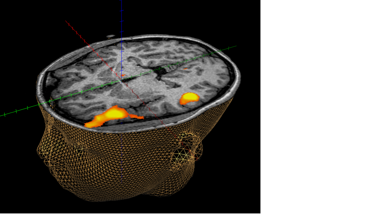
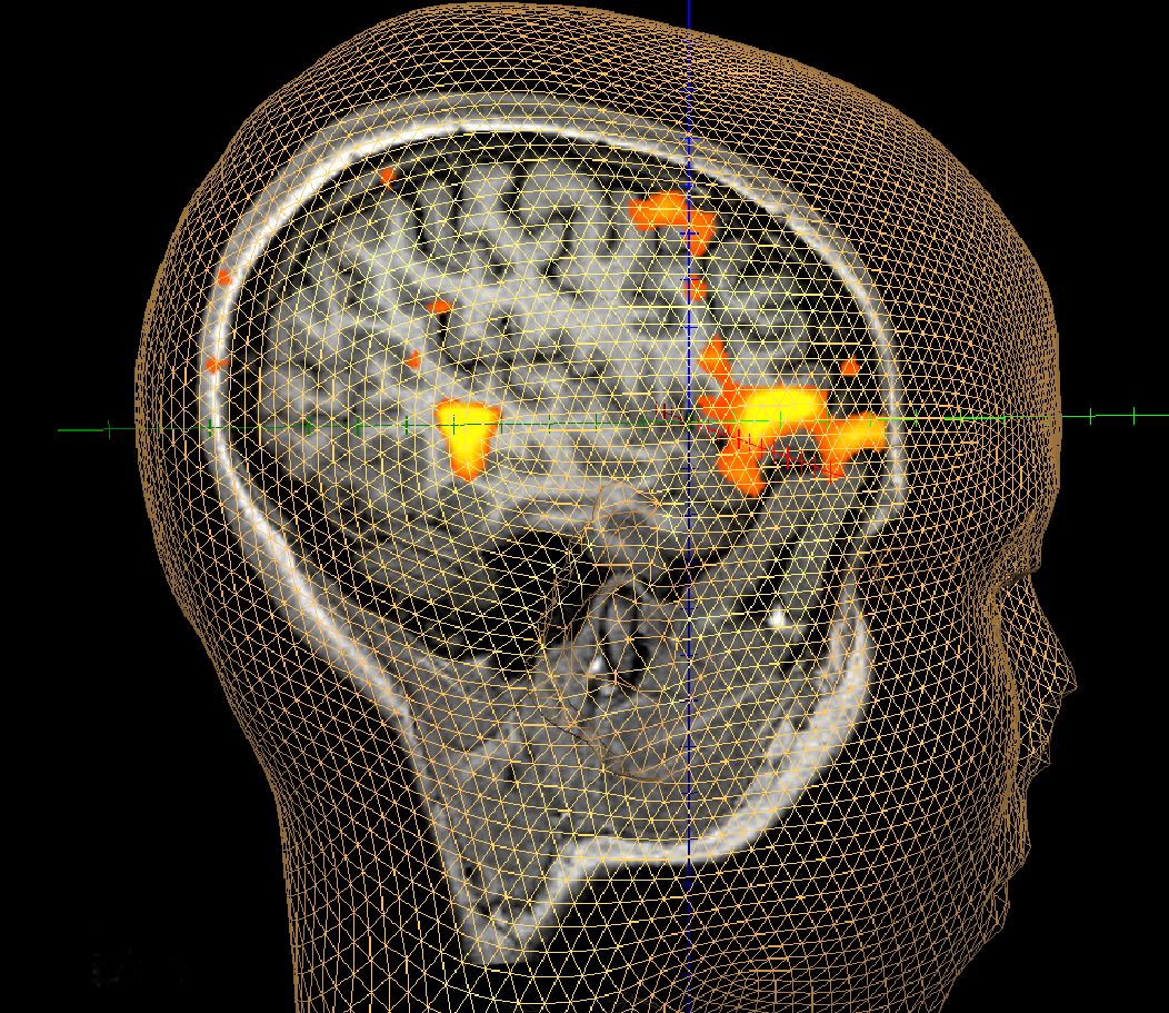
The fundamental difference that EZfMRI offers is a simple paradigm delivery system through the existing auditory communication system of the MRI scanner. Since it is used every day at the imaging site, it is tested and known to be working all the time. There is no complicated additional hardware to maintain or purchase so any site can afford to offer fMRI studies. EZfMRI uses simple robust paradigms that have been clinically tested at our academic partner’s facility in the inpatient and outpatient setting. These tasks have been vetted on clinical patients so the customer can be confident that they are offering the best fMRI exam for their patients. Finally, the EZfMRI product will come complete with automated post-processing protocols to create the functional maps correctly. The MR technologist and Radiologist can take advantage of the built-in software of their MRI scanner to generate the functional maps without having to export the data to a third party system, process the data and then import the data back into PACS. The activation maps will be automatically generated with the EZfMRI protocols and stored with the patient’s study for easy interpretation by the Radiologist. More importantly, they can be evaluated while the patient is still on the table in case additional studies are required.
The auditory based paradigms and post-processing protocols delivered by EZfMRI will provide a cost effective (~$10k range), highly efficient method to conduct fMRI studies at any MR imaging center using any vendor scanner with the appropriate software license. The system will exploit the auditory system, already employed by the site to play music, to deliver instructions and robust paradigms that are easy to follow and clinically tested. Finally, the implementation of post-processing on the scanner hardware eliminates any additional devices that require support. EZfMRI provides an economical solution for conducting high quality clinical fMRI studies.